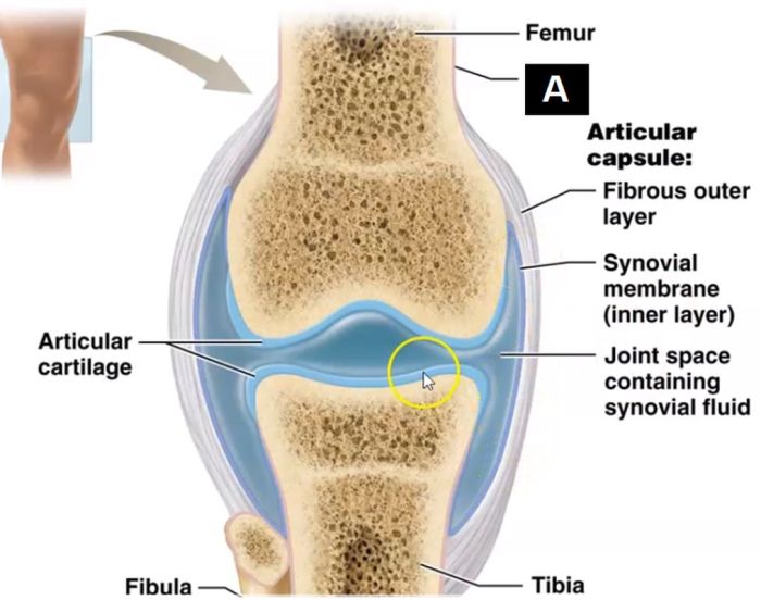
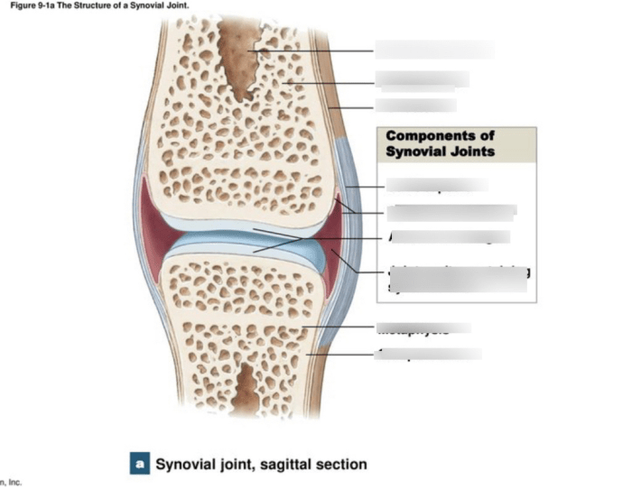
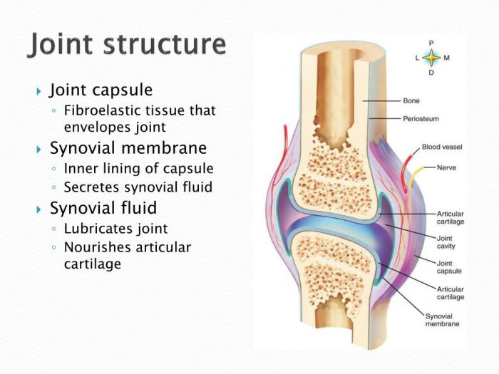
Embark on a comprehensive exploration of the art-labeling activity structure of a typical synovial joint. This detailed guide unveils the intricate anatomy, function, and interconnections of this vital bodily structure, providing a profound understanding of its significance in human movement and well-being.
Delve into the intricacies of synovial joint anatomy, articular cartilage, synovial membrane, joint capsule, ligaments, tendons, joint innervation, and joint blood supply. Each component is meticulously described, illustrated, and analyzed to provide a holistic perspective on the joint’s structure and function.
Synovial Joint Anatomy: Art-labeling Activity Structure Of A Typical Synovial Joint

Synovial joints are the most common type of joint in the human body. They are characterized by the presence of a joint cavity filled with synovial fluid, which lubricates and nourishes the joint. Synovial joints allow for a wide range of motion, including flexion, extension, rotation, and abduction.
Types of Synovial Joints
- Hinge jointsallow for flexion and extension in one plane, such as the knee and elbow joints.
- Pivot jointsallow for rotation around a single axis, such as the atlanto-axial joint in the neck.
- Condyloid jointsallow for flexion, extension, abduction, and adduction, such as the wrist and ankle joints.
- Saddle jointsallow for flexion, extension, abduction, and adduction in two perpendicular planes, such as the thumb joint.
- Ball-and-socket jointsallow for the widest range of motion, including flexion, extension, rotation, abduction, and adduction, such as the hip and shoulder joints.
Articular Cartilage
Articular cartilage is a thin layer of hyaline cartilage that covers the ends of bones at synovial joints. It is responsible for reducing friction and providing a smooth surface for joint movement.
Articular cartilage is composed of chondrocytes, which are specialized cells that produce the extracellular matrix. The extracellular matrix is made up of collagen, proteoglycans, and water. Collagen provides strength and flexibility to the cartilage, while proteoglycans attract water, which gives the cartilage its cushioning properties.
Articular cartilage is avascular, which means that it does not have any blood vessels. As a result, it is very slow to heal if it is damaged.
Synovial Membrane
The synovial membrane is a thin layer of tissue that lines the joint cavity. It is responsible for producing synovial fluid, which lubricates and nourishes the joint.
The synovial membrane is composed of two layers: the intima and the subintima. The intima is the inner layer and is made up of synovial cells. Synovial cells produce synovial fluid, which is a clear, viscous fluid that contains hyaluronic acid, proteins, and cells.
The subintima is the outer layer of the synovial membrane and is made up of connective tissue. The subintima contains blood vessels, nerves, and lymphatics.
Joint Capsule, Art-labeling activity structure of a typical synovial joint
The joint capsule is a tough, fibrous membrane that surrounds the synovial joint. It is responsible for providing stability and support to the joint.
The joint capsule is composed of two layers: the outer fibrous layer and the inner synovial layer. The outer fibrous layer is made up of collagen fibers, which provide strength to the capsule. The inner synovial layer is made up of synovial cells, which produce synovial fluid.
The joint capsule is attached to the bones at the joint by ligaments. Ligaments are tough, fibrous bands of tissue that help to stabilize the joint.
Ligaments
Ligaments are tough, fibrous bands of tissue that connect bones to other bones. They are responsible for providing stability and support to joints.
There are two main types of ligaments: extrinsic and intrinsic ligaments. Extrinsic ligaments are located outside the joint capsule, while intrinsic ligaments are located within the joint capsule.
Extrinsic ligaments are usually stronger than intrinsic ligaments. They provide stability to the joint in all directions.
Intrinsic ligaments are usually weaker than extrinsic ligaments. They provide stability to the joint in specific directions.
Tendons
Tendons are tough, fibrous bands of tissue that connect muscles to bones. They are responsible for transmitting the force of muscle contractions to bones.
Tendons are composed of collagen fibers, which are arranged in a parallel fashion. This gives tendons their strength and flexibility.
Tendons are usually located outside the joint capsule. However, some tendons may pass through the joint capsule to attach to bones within the joint.
Joint Innervation
Synovial joints are innervated by both sensory and motor nerves. Sensory nerves provide sensation to the joint, while motor nerves control the muscles that move the joint.
The sensory nerves that innervate synovial joints are located in the joint capsule, synovial membrane, and ligaments. These nerves provide information about the position of the joint, the amount of force that is being applied to the joint, and the presence of pain.
The motor nerves that innervate synovial joints are located in the muscles that move the joint. These nerves control the contraction and relaxation of the muscles, which allows the joint to move.
Joint Blood Supply
Synovial joints are supplied with blood by arteries and veins. The arteries supply the joint with oxygen and nutrients, while the veins remove waste products from the joint.
The arteries that supply synovial joints are located in the joint capsule and synovial membrane. The veins that drain synovial joints are located in the joint capsule and synovial membrane.
The blood supply to synovial joints is important for the health of the joint. It provides the joint with the oxygen and nutrients that it needs to function properly, and it removes waste products from the joint.
Q&A
What is the primary function of articular cartilage in synovial joints?
Articular cartilage provides a smooth, low-friction surface for joint movement, reducing wear and tear and facilitating gliding motions.
How does the synovial membrane contribute to joint lubrication?
The synovial membrane secretes synovial fluid, which acts as a lubricant, reducing friction and providing nutrients to the joint.
What is the role of ligaments in joint stability?
Ligaments are tough, fibrous bands of connective tissue that connect bones across joints, providing stability and preventing excessive movement.


